Home » Without Label » Labelled Muscular System Front And Back / Labelled Muscular System Front And Back / Muscles_anterior ... - Labelled muscular system front and back / muscles, connected to bones or internal organs and blood vessels, are in charge for movement.
Labelled Muscular System Front And Back / Labelled Muscular System Front And Back / Muscles_anterior ... - Labelled muscular system front and back / muscles, connected to bones or internal organs and blood vessels, are in charge for movement.
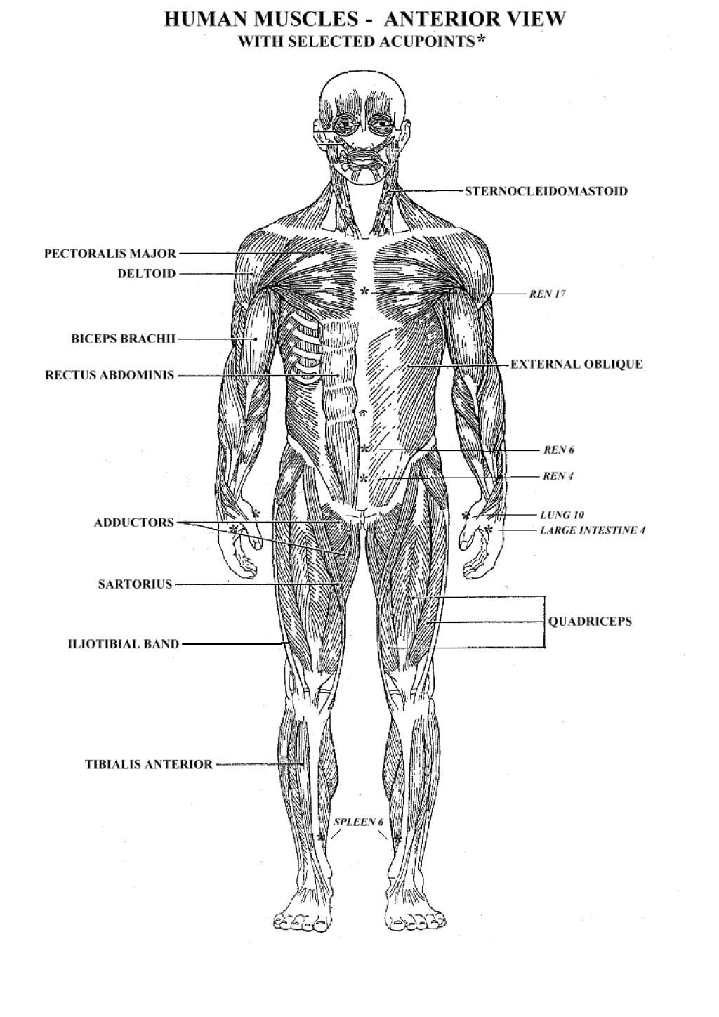
Labelled Muscular System Front And Back / Labelled Muscular System Front And Back / Muscles_anterior ... - Labelled muscular system front and back / muscles, connected to bones or internal organs and blood vessels, are in charge for movement.. Human muscle system, the muscles of the human body that work the skeletal system, that are under voluntary control, and that are concerned with movement, posture, and balance.broadly considered, human muscle—like the muscles of all vertebrates—is often divided into striated muscle (or skeletal muscle), smooth muscle, and cardiac muscle.smooth muscle is under involuntary control and is. In this image, you will find frontalis, orbicularis oculi, zygomaticus, masseter, orbicularis oris, sternocleidomasteoid, deltoid, pectoralis major, biceps brachii, iliopsoas, adductor longus, gastrocnemius. Back pain is one of the most common kinds of pain for adults, and muscle strains are the most common type of back pain. We are pleased to provide you with the picture named anatomy of back muscles diagram. The muscular system the human muscular system vector illustration, front and rear view muscle anatomy with labels stock illustrations labeled anatomy chart of neck and shoulder muscles on black background labeled human anatomy diagram of man's neck and shoulder muscles in an anterior view on a black background.
Use your front view and back view diagrams to label these muscles gluteus medius label:place the letter next to the name. For more anatomy content please follow us and visit our website: The front fibers flex the arm and the middle fibers help abduct the arm (bring the arm away from the body). Other muscles are small and cover much less space. Muscles are the only tissue in the body that has the ability to contract and therefore move the other parts of the body.
Labelled Muscular System Front And Back : Google Image ... from i.pinimg.com Labelled muscular system front and back / muscles, connected to bones or internal organs and blood vessels, are in charge for movement. The maintenance of posture and body. Muscles in the torso protect the internal organs at the front, sides, and back of the body. The muscles that make up the quadriceps are the strongest and leanest of all muscles in the body. Muscle labeling worksheet kids health. Vector illustration informative medical scheme. Vector flat color illustration set. Muscle or ligament strains can occur from repeated use of the muscles, or from improperly or awkwardly lifting heavy objects.
Free anatomy quiz the muscular system section back muscle diagram labeled best 15 bright human body muscles muscular system rear labeled body part chart removable wall the muscular system these are the major muscles of the body
Male shoulder and chest muscles labeled chart on black labeled human anatomy diagram of male shoulder, biceps, arm, and chest muscles frontal anterior view on a black background. The muscular system is an organ system consisting of skeletal, smooth and cardiac muscles. Posterior (back) fibers help to extend the arm. The muscular system is an organ system consisting of skeletal. The back's muscles start at the top of the back (named the cervical vertebrae) and go to the tailbone (also named the coccyx). Related posts of muscles labeled front and back. Muscles labeled front and back find out more about muscles labeled front and back. Flat medical scheme poster of training healthcare gym ,. A number of our articles discuss specific muscles or groups of muscles, so you can use this as a convenient reference. Labelled muscular system front and back / muscles, connected to bones or internal organs and blood vessels, are in charge for movement. Human body anatomy male man , front and back muscular system of muscles. This large muscle in the back. To see a muscular system picture from the anterior (front) view click here.
Posterior (back) fibers help to extend the arm. Back pain is one of the most common kinds of pain for adults, and muscle strains are the most common type of back pain. Muscle anatomy with labels stock. Related posts of muscles labeled front and back. Human body muscle system, the muscles of the human body that work the skeletal system, that are under voluntary control, and that are concerned with movement, posture, and balance.
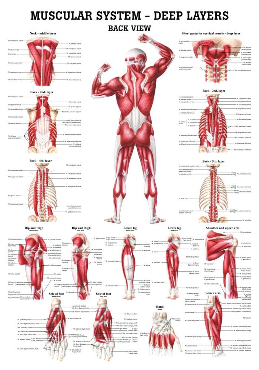
Human Muscular Systems-Deep Layers of the Back Poster ... from cdn11.bigcommerce.com Use your front view and back view diagrams to label these muscles gluteus medius label:place the letter next to the name. Muscles labeled front and back find out more about muscles labeled front and back. Muscle anatomy with labels stock. Other muscles are small and cover much less space. The muscle fibers' highly specialized structure enables the muscles to relax and contract to produce movement. We hope this picture anatomy of back muscles diagram can help you study and research. We are pleased to provide you with the picture named anatomy of back muscles diagram. Learn about and revise the muscular system with this bbc bitesize gcse pe (edexcel) study guide.
This rotator cuff muscle helps with the raising and lowering of the upper arm.;
Muscle or ligament strains can occur from repeated use of the muscles, or from improperly or awkwardly lifting heavy objects. Unlabeled muscular system front and back these pictures of this page are about:muscular system labeled back. Muscle diagram labeled front and back, muscle system labelling (front and back), muscular system labeled front and back, human muscles, muscle diagram labeled front and back, muscle system labelling (front and back), muscular system labeled front and back. Posterior (back) fibers help to extend the arm. The back's muscles start at the top of the back (named the cervical vertebrae) and go to the tailbone (also named the coccyx). In this image, you will find frontalis, orbicularis oculi, zygomaticus, masseter, orbicularis oris, sternocleidomasteoid, deltoid, pectoralis major, biceps brachii, iliopsoas, adductor longus, gastrocnemius. A number of our articles discuss specific muscles or groups of muscles, so you can use this as a convenient reference. Human body muscle system, the muscles of the human body that work the skeletal system, that are under voluntary control, and that are concerned with movement, posture, and balance. To see a muscular system picture from the anterior (front) view click here. Muscles labeled front and back muscular anatomy simple detail ideas free cool example gallery · august 20, 2016 related posts of muscles labeled front and back We are pleased to provide you with the picture named anatomy of back muscles diagram. Labelled muscular system front and back attached to the bones creatine phosphate donates its phosphate group to adp to turn it back into atp in order to. The muscles that make up the quadriceps are the strongest and leanest of all muscles in the body.
Vector flat color illustration set. Other muscles are small and cover much less space. Muscles labeled front and back muscular anatomy simple detail ideas free cool example gallery · august 20, 2016 related posts of muscles labeled front and back In this image, you will find frontalis, orbicularis oculi, zygomaticus, masseter, orbicularis oris, sternocleidomasteoid, deltoid, pectoralis major, biceps brachii, iliopsoas, adductor longus, gastrocnemius. It is controlled by the axillary nerve.
The Muscular System Coloring Pages - Coloring Home from coloringhome.com For more anatomy content please follow us and visit our website: The muscles, bones, ligaments, and tendons in the back can all be injured and cause back pain. Related to the function of movement is the muscular system's second function: Other muscles are small and cover much less space. Posterior (back) fibers help to extend the arm. This muscular system diagram shows the major muscle groups from the back or posterior view. This labeled human muscular system chart illustrates the major muscle groups in the back (posterior) view and the front (anterior) view. The muscular system is an organ system consisting of skeletal, smooth and cardiac muscles.
The muscular system is an organ system consisting of skeletal, smooth and cardiac muscles.
Other muscles that aid in shoulder movement include: The muscular system is an organ system consisting of skeletal. Muscular system physiology function of muscle tissue. Vector flat color illustration set. The muscles that make up the quadriceps are the strongest and leanest of all muscles in the body. Wide selection, fast delivery and awesome service. Muscles are the only tissue in the body that has the ability to contract and therefore move the other parts of the body. Related posts of muscles labeled front and back. The muscular system the human muscular system vector illustration, front and rear view muscle anatomy with labels stock illustrations labeled anatomy chart of neck and shoulder muscles on black background labeled human anatomy diagram of man's neck and shoulder muscles in an anterior view on a black background. We are pleased to provide you with the picture named anatomy of back muscles diagram. The main function of the muscular system is movement. The muscular system is an organ system consisting of skeletal, smooth and cardiac muscles. Some of these muscles are quite large and cover broad areas.